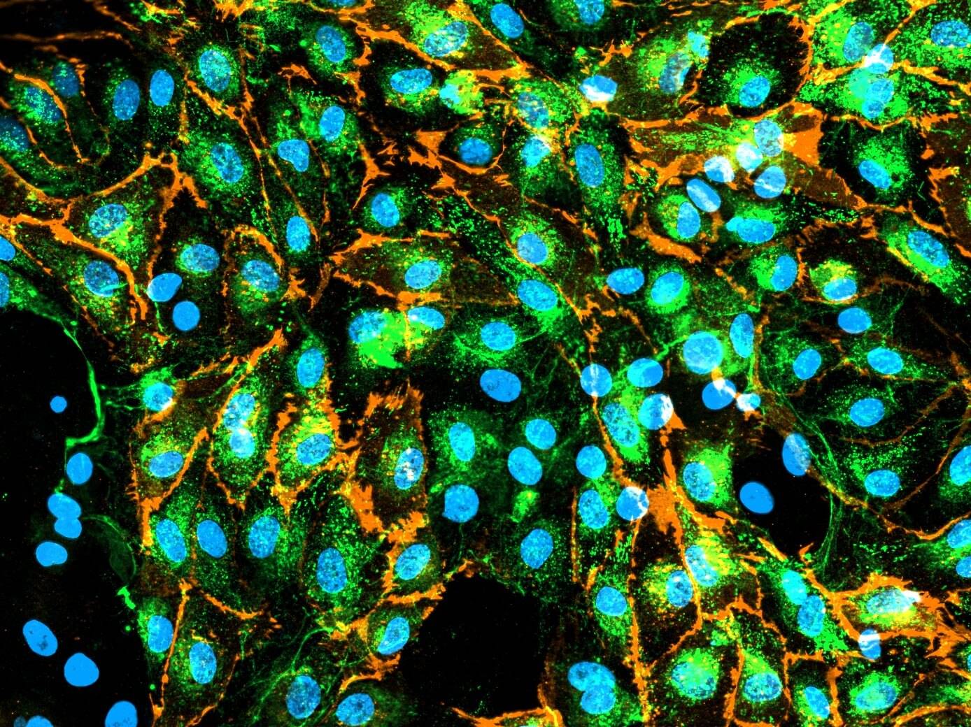
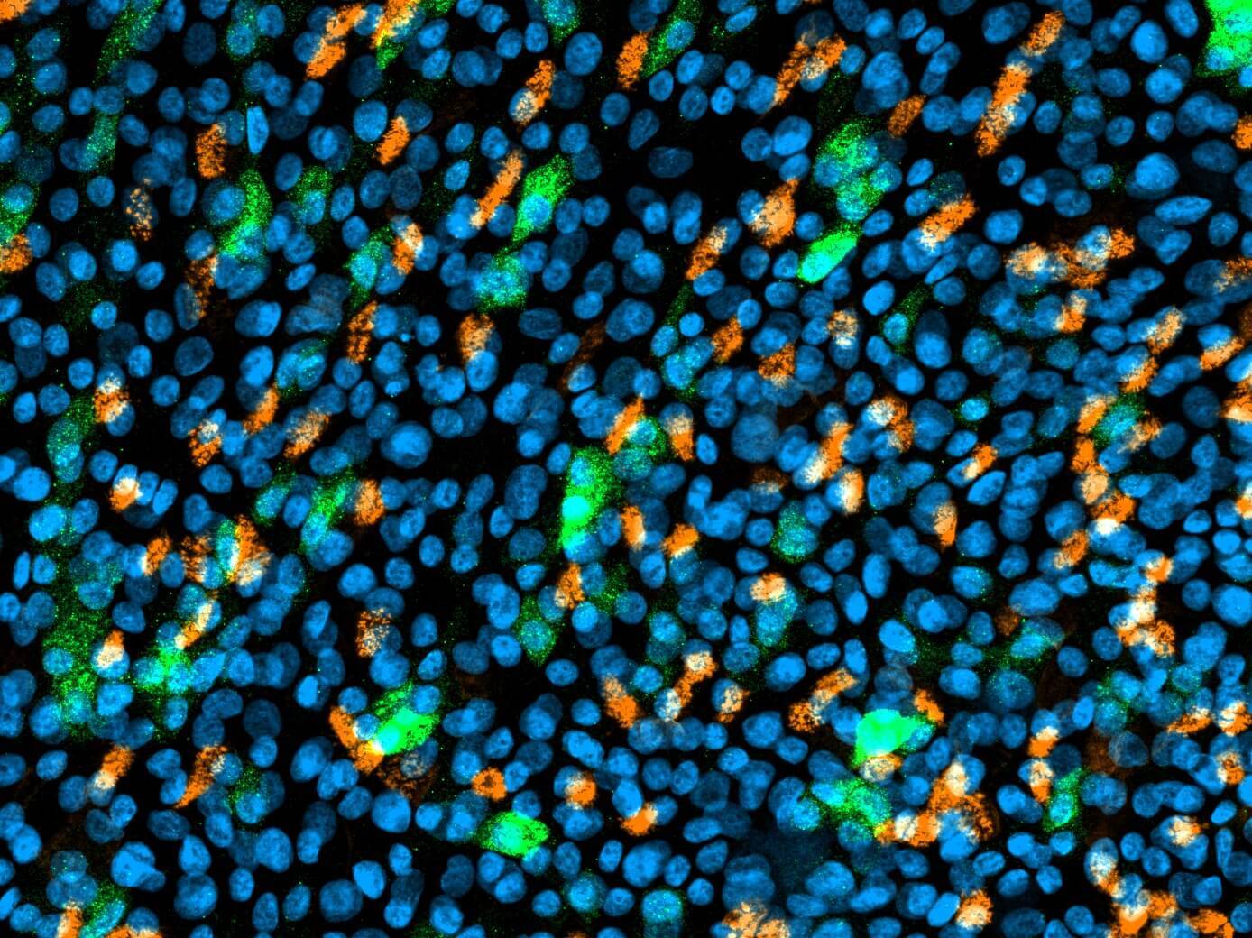
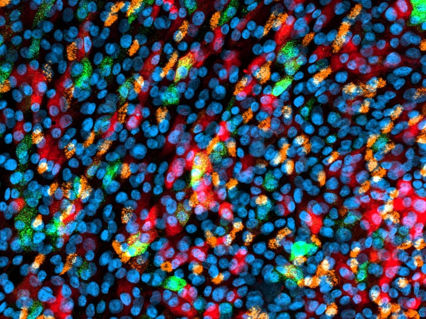
D42 Lung-on-Chip: Endothelial cells comprise the vasculature of our Lung-on-Chip model. Endothelial cells express von Willebrand factor (vWf) stained in green and CD31 stained in orange. vWf plays a major role in platelet adhesion and in protective complex formation with factor VIII. CD31 mediates mechano-sensing of biomechanical stimuli and leukocyte adhesion to the vessel wall.

The Dynamic42 Lung-on-Chip can be used to evaluate uptake and mechanistical features of pre-clinical drug candidates and chemicals that follow a respiratory delivery via the lung epithelium. It further qualifies to investigate air-borne infectious diseases.
We use our perfusable Dynamic42 biochip platform to provide an innovative concept to setup in vivo-like microenvironments of the human lung by reconstructing perfusable vascular tissue in combination with lung epithelium exposed to air.
Specifications
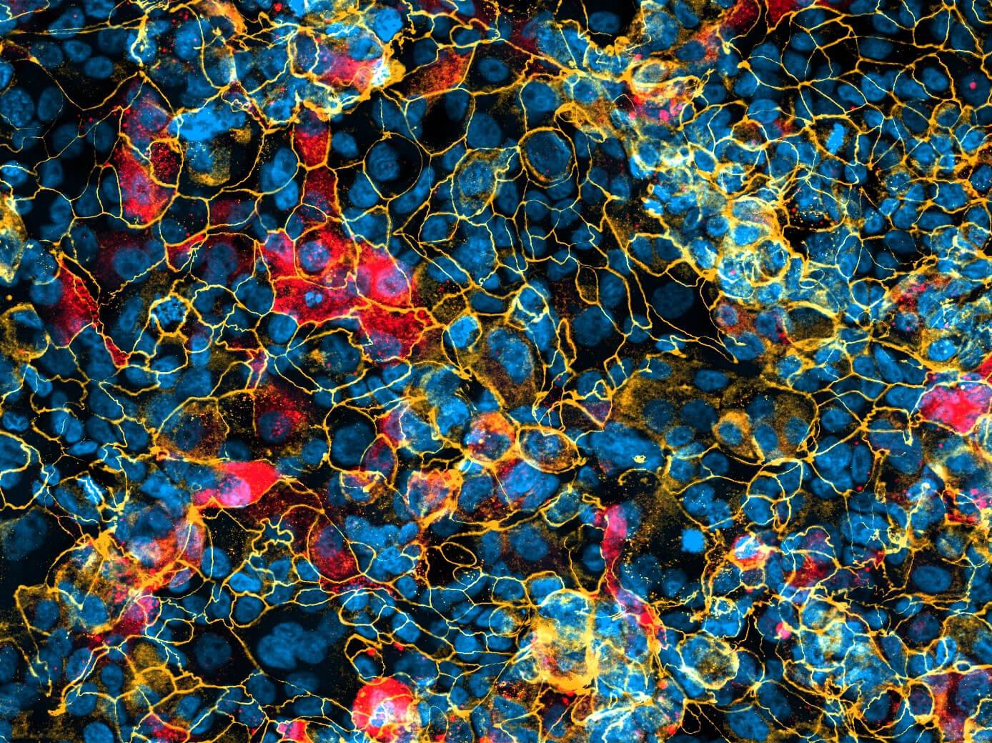
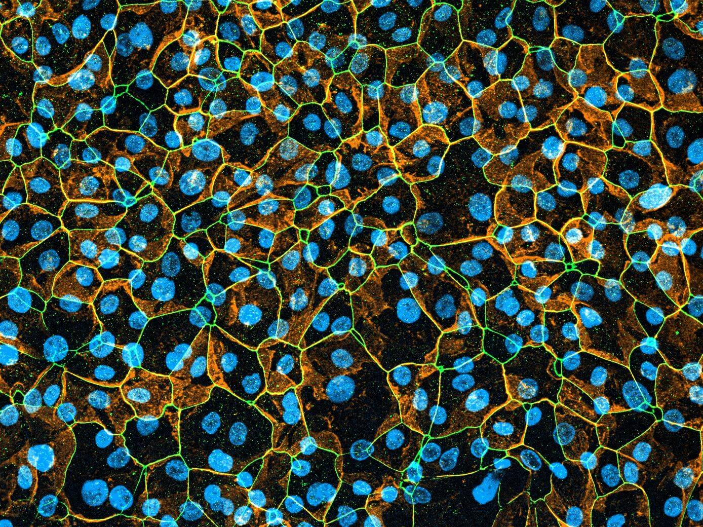
The Dynamic42 Lung-on-Chip comprises two compartments: a vascular compartment and an alveolar compartment. The vasculature is formed by an endothelial lining. The alveolar compartment comprises lung epithelial cells and tissue-resident alveolar macrophages. The alveolar tissue is exposed to air and nourished from the vasculature below (air-liquid-interface culture).
The Dynamic42 Lung-on-Chip can be operated with various cell sources. Please get in contact for further details.


Macrophage

Epithelial Cell

Endothelial Cell
Two models can be operated in parallel on one Dynamic42 biochip.
The model has been tested in the following Dynamic42 biochips: BC001 and BC002.
Characteristics

Enhanced marker expression

Intense cell-cell communication

Physiologic biomechanical stimulation via flow
Secretory function

Immunocompetence
The Dynamic42 Lung-on-Chip can be operated up to 14 days with high vitality, stable barrier function and stable marker expression.


Applications
- Toxicity profiling
- Uptake / Transport studies
- Inflammatory Diseases
- Infectious Diseases